Replica creation service for autologous tooth transplantation
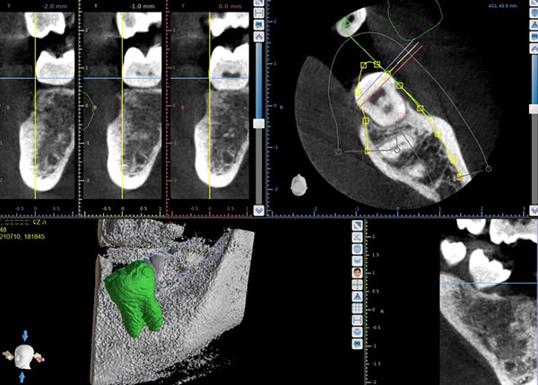
Created from DICOM data
Dental Prosthesis Modeling Service
simulation model
$85(Shipping cost included)
Please send DICOM data to shibuya8020@gmail.com.For CT imaging, it is sufficient if the field of view (FOV) is narrowed and the implanted tooth is shown. It doesn't matter if it's not full chin.
↓↓↓ Enter the CT data here and we will start production
Tel +81 48-961-8151
To increase the success rate of autologous tooth transplantation.
- To extract and immediately implant the transplanted tooth into the prepared recipient site.
- To avoid damaging the periodontal tissues (periodontal ligament) surrounding the tooth root.
- Young individuals (preferably below 40 years of age, although successful cases have been observed in individuals up to 70 years of age).
- To ensure that the transplanted tooth has appropriate bone width.
One of the conditions to increase the success rate of autologous tooth transplantation is time. It is crucial to implant the extracted tooth (transplanted tooth) into the alveolar bone (recipient site) as quickly as possible without causing damage. By using 3D replicas, it becomes possible to immediately perform the implantation.
Without using 3D replicas, the process of implanting the transplanted tooth into the alveolar bone after extraction involves carefully shaping the recipient site while positioning the transplanted tooth. This method takes more time and physically damages the transplanted tooth (periodontal ligament).
By using 3D replicas of teeth, it becomes possible to perform preoperative simulations of tooth transplantation. This allows for a better understanding of the shape of the tooth, confirmation of jawbone reduction techniques, and assessment of occlusion. Tasks that could only be performed after the extraction of the transplanted tooth can now be conducted in advance.
Preserving the periodontal tissues, especially the remnants of Malassez's epithelium, without causing damage, is crucial in autogenous tooth transplantation and replantation treatment.
In immature teeth with incomplete root formation, the presence of Hertwig's epithelial sheath at the apical region is important for the healing of the dental pulp. Therefore, it is crucial to avoid damaging the periapical tissues in order to preserve the integrity of the root apex.
To increase the success rate of autogenous tooth transplantation
One of the important factors for increasing the success rate of autogenous tooth transplantation is to promptly implant the extracted tooth (transplant tooth) into the alveolar bone (transplant bed). By using a 3D replica, the morphology of the transplant tooth can be confirmed, enabling the preparation of the transplant bed in advance.
Case Presentation
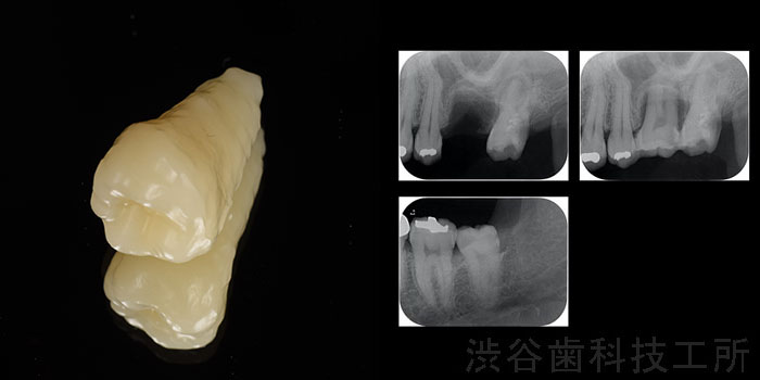
Case 1
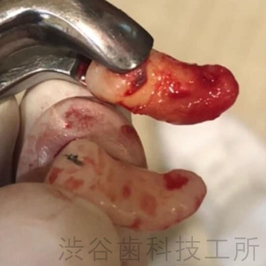
In order to create a smooth and accurate transplant bed (socket on the recipient side) tailored to the dental implant, the morphology of the dental implant is prepared in advance by replacing it with PMMA (resin) based on CT data
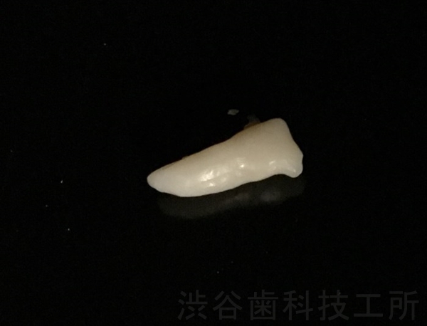
In this case, a donor replica was created for the transplantation of the peg-shaped tooth (tooth number 8) to tooth number 4.
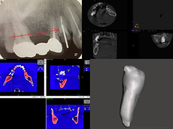
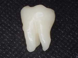
We extract dental data, not only including the morphology of the tooth root but also the crown portion.
compared them
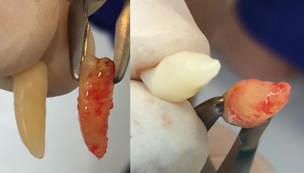
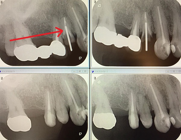
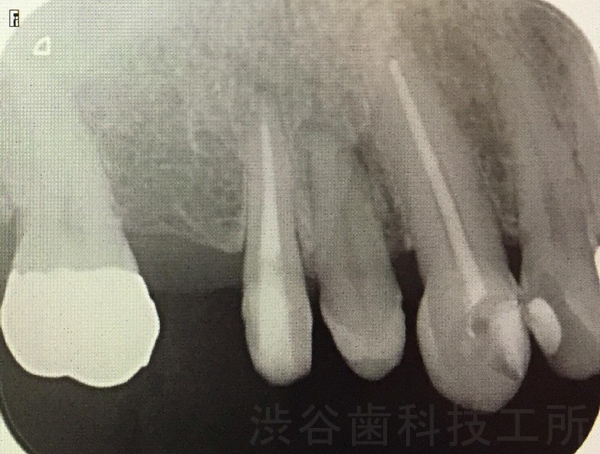
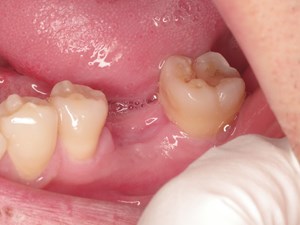
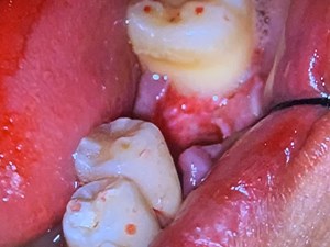
After the surgery
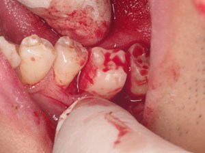
Case 2


We proceed with the formation of the transplant bed while confirming the fit using a 3D replica of the dental implant before extracting the donor tooth.
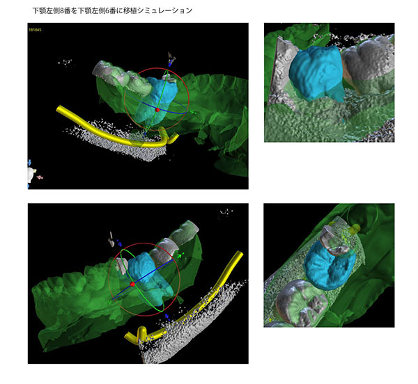
Case 3
making theeth
Transplantation simulation of artificial tooth root by using the 3D printing model
autotransplantation of teeth with 3D printed surgical guides
As a valuable method for restoring missing teeth, autotransplantation of teeth has been used in clinical practice for over 60 years
Dental autotransplan
autotransplantation
3D donor replica root